

XERA Medical Systems and Technology actively participated in the SPIE Medical Imaging Conference 2026 in Vancouver, presenting its ongoing research in advanced X-ray technologies.
XERA focuses on research and development in detectors, sensors, and detection systems, combining device physics, hardware engineering, and intelligent imaging solutions. The company continues to expand its work in these areas and contribute to the medical imaging community through both scientific research and practical system development.
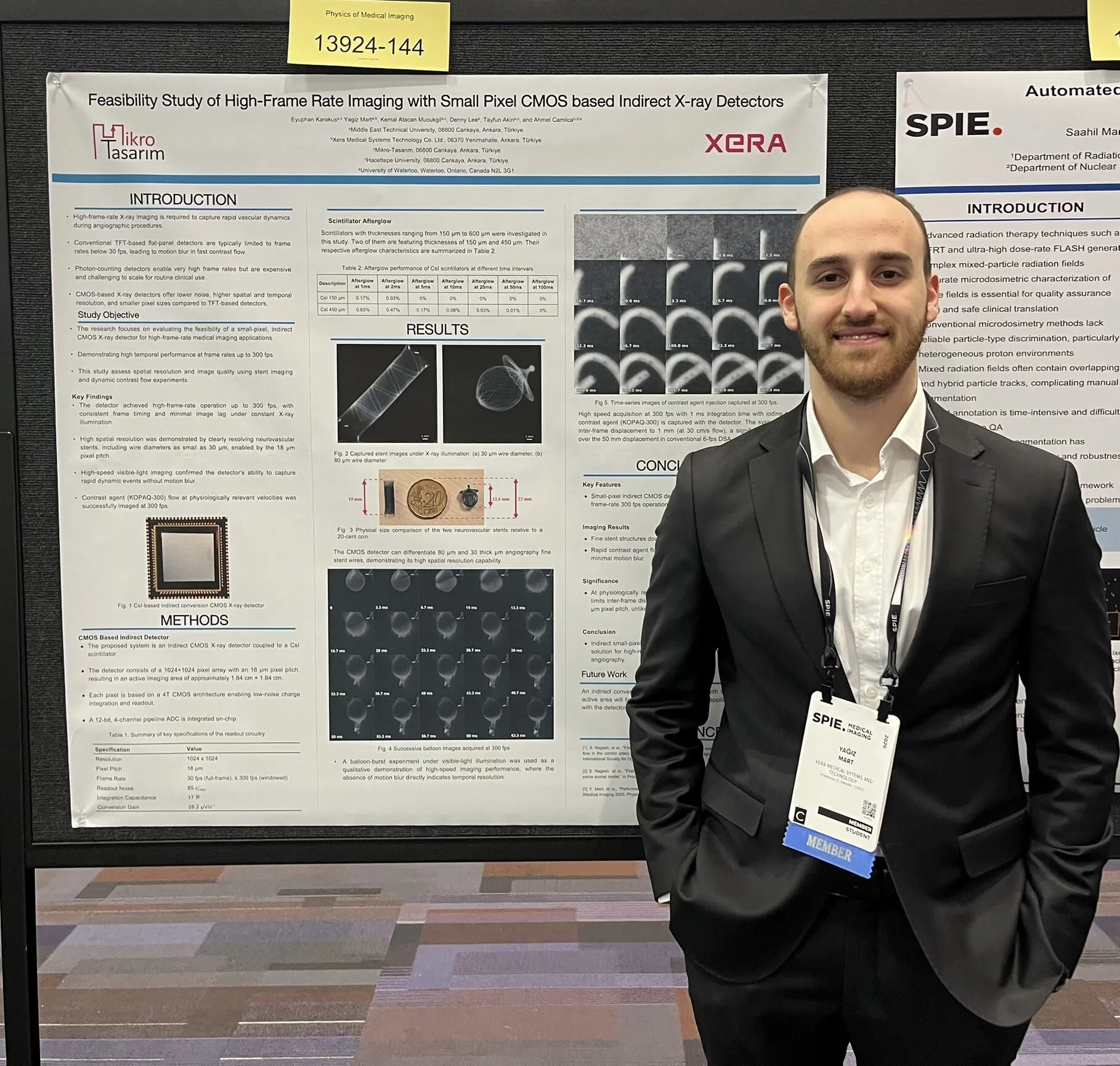
High-Frame-Rate CMOS X-Ray Detector Study
One of the presenters, Yağız Mart, serves as an R&D Engineer at XERA, focusing on X-ray detector technologies and performance optimization, while also pursuing an M.Sc. degree in the Electronics Department at Middle East Technical University (METU).
He presented the work titled “Feasibility Study of High-Frame-Rate Imaging with Small Pixel CMOS-Based Indirect X-Ray Detectors” in the poster session.
The study investigated the feasibility of using an 18 µm pixel CMOS detector architecture for dynamic imaging applications such as angiography. The prototype system is a 1024 × 1024 CsI-coupled CMOS detector featuring:
• Up to 300 fps operation in ROI mode
• 4.7 lp/mm MTF spatial performance
• ~85 e⁻ RMS input-referred noise
• Negligible image lag at high frame rates
Temporal and spatial performance were validated through dynamic contrast-agent flow imaging and neurovascular stent visualization. The results demonstrate that indirect CMOS technology can achieve high temporal resolution while small pixel sizes enable superior spatial resolution, offering a clinically practical alternative for high-speed X-ray imaging.
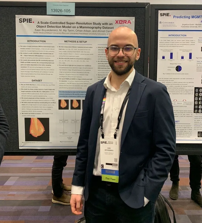
AI-Based Super-Resolution Study in Mammography
Kaan Büyükdemirci, who leads XERA’s AI and software development efforts while continuing his master’s studies at Bilkent University, presented the work titled “A Scale-Controlled Super-Resolution Study with YOLOv11 on VinDr-Mammo” in the poster session.
In this study, we conducted a scale-controlled and task-oriented evaluation using the VinDr-Mammo dataset. We generated low-resolution variants (128, 256, 512, and 1024 pixels) and trained independent YOLOv11 object detectors at each resolution. We then evaluated SR-to-object-detection (SR → OD) pipelines using 2× and 4× super-resolution and compared them against bilinear up-sampling and native-resolution baselines.
To reduce domain variability, we restricted the dataset to Siemens Mammomat cases with mass annotations and created dual-view (CC + MLO) composite images on a square canvas, enabling the detector to leverage cross-view context.
Our findings show that while super-resolution consistently improves PSNR and SSIM compared to bilinear interpolation, task-level performance gains depend strongly on the native sampling resolution.
• At 128 px, SR does not outperform the native detector.
• From 256 px, 4× SR improves mAP from 0.493 (native) and 0.526 (bilinear) to 0.573.
• From 512 px, 2× SR improves mAP from 0.503 (native) and 0.574 (bilinear) to 0.592.
These results suggest that super-resolution becomes beneficial once lesions occupy a minimal spatial footprint in the image. Interestingly, SR-generated inputs may align more effectively with a detector’s learned feature representations than those obtained through raw higher-resolution training.
We believe this work contributes to a more realistic understanding of when super-resolution is clinically meaningful in mammography AI pipelines.
Amorphous Selenium Drift Detector Architecture
Another SPIE presentation was delivered in the oral session by Mohammad Ala A. Mohajerzadeh, a Ph.D. candidate at the University of Waterloo.
He presented the work titled “Unipolar Charge Sensing Drift Detector Using Amorphous Selenium for Large-Area X-Ray Imaging Applications.”
This study introduced a novel large-area amorphous selenium (α-Se) drift detector architecture based on field-shaping strips inspired by the Frisch grid concept. The design addresses a fundamental limitation of conventional α-Se detectors, the low mobility and higher trapping probability of electrons, which negatively affect signal rise time and energy resolution.
By engineering semi-drift electric fields within the selenium bulk, the field-shaping strips enable unipolar charge sensing, preferentially enhancing hole collection while suppressing the influence of slower electrons. Since holes are the faster charge carriers in α-Se, this approach:
• Improves charge collection efficiency
• Reduces signal rise time
• Enhances photocurrent response
• Mitigates energy resolution degradation caused by electron trapping
Additionally, a novel hole-blocking layer was implemented to suppress dark current, a major contributor to noise in large-area photoconductive detectors. The combined effect of enhanced hole transport and reduced dark current is expected to significantly improve SNR and overall detector performance.
Initial validation using blue LED illumination demonstrated promising hole-drift behavior in the field-shaped configuration. X-ray characterization with proper collimation is planned as the next stage toward large-area imaging applications.
The conference provided an excellent opportunity to connect with field experts, academics, and students. Strong professional relationships were established, opening the door to new collaborations. It was also valuable to present our work to the scientific community and actively contribute to ongoing research efforts.
XERA looks forward to continuing its research in detector and imaging technologies and further strengthening its international collaborations in the coming years. The company plans to continue participating in SPIE Medical Imaging annually.
- Prev
-
List

